Anatomical illustrations demonstrating the facial transplant procedure.
The first facial tissue transplant surgery of Finland and the Nordic Countries was carried out in Helsinki in 2016. I collaborated with the leading surgeon Patrik Lassus and another forensic artist/anatomist Lydia Carline to create anatomical illustrations to explain the procedure to the general public and professionals in healthcare fields. We created over 30 illustrations altogether, some of which were widely distributed in the Finnish media. Here are some examples.
A few media links (in Finnish but with illustrations):
http://yle.fi/uutiset/3-8690134
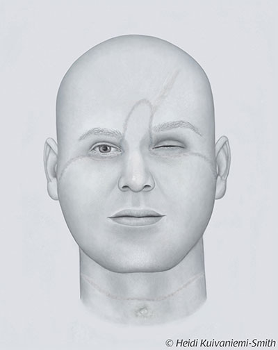
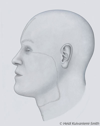
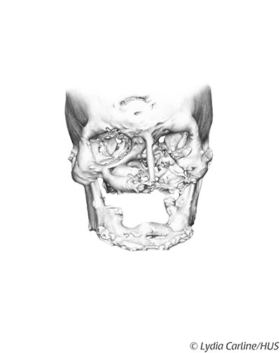
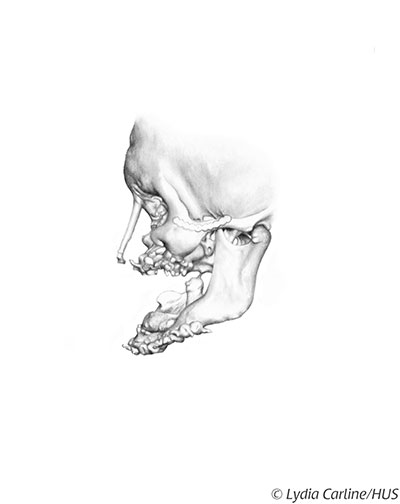
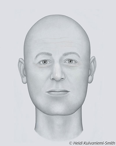
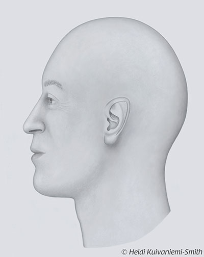
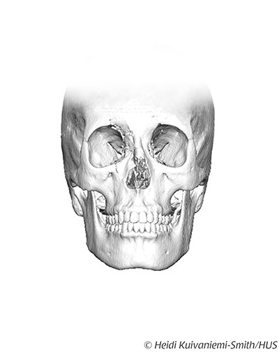
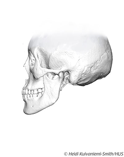
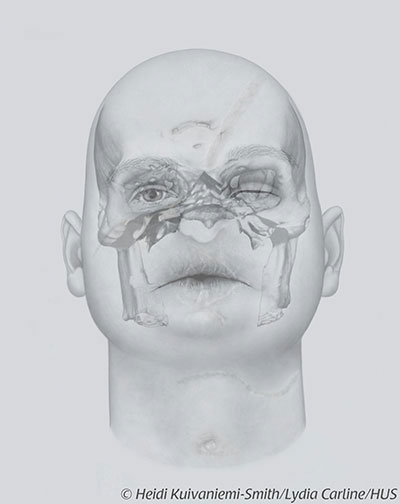
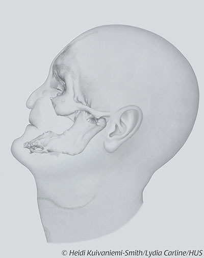
Facial illustrations of the patient before the surgery. Below illustrations of the patient’s damaged skull.
Facial illustrations of the braindead donor (what they would have looked like alive and well) and below the corresponding skull.
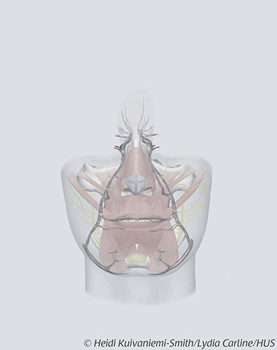
The part of the face that was used as a transplant highlighted in the donor’s face.
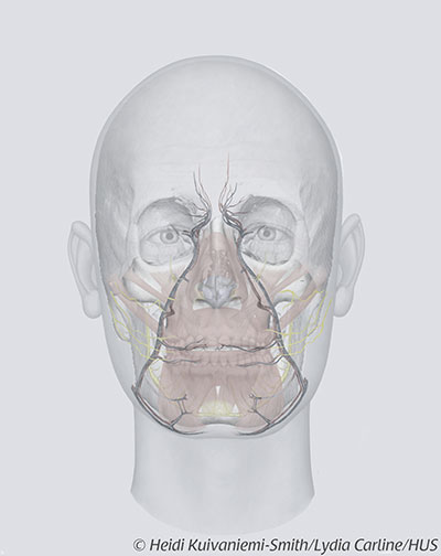
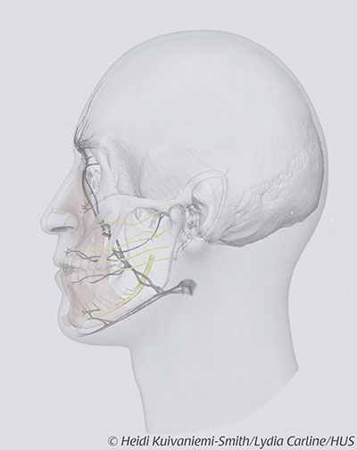
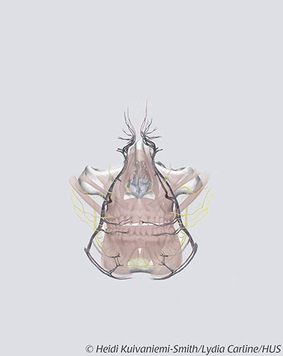
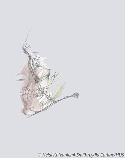
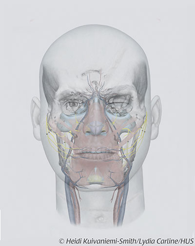
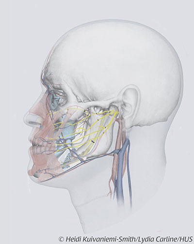
The transplant part containing facial bones, facial muscles, blood vessels and nerves. All these layers including skin were transplanted onto the patient’s face.
Illustrations showing the damaged bones removed from the patient’s midface.
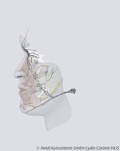
Illustrations demonstrating the transplant having been attached to the patient’s face.
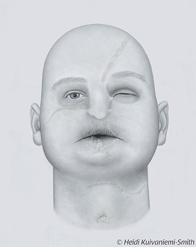
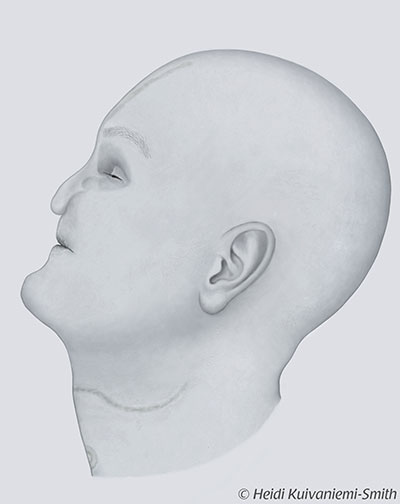
The patient’s new face after facial transplant surgery.